Digital radiography has transformed how dental teams capture and use images to evaluate oral health. By replacing traditional film with sensitive electronic sensors and computer processing, modern x-ray systems deliver clearer images in seconds while streamlining workflows in the operatory and the office computer. The result is a smoother visit for patients and more efficient, evidence-based decision making for clinicians. Below are practical explanations of how digital radiography works and why it matters to routine care and more complex treatment planning.
At its core, digital radiography converts x-ray exposure into a digital file that can be viewed, manipulated, and stored on a computer. Instead of waiting for film to be developed, clinicians see high-resolution images almost immediately on a monitor. This immediacy helps patients and providers review findings together during the appointment, improving communication and helping people understand the condition of their teeth and supporting structures.
Digital sensors come in several shapes and sizes to fit comfortably inside the mouth or to capture larger areas from outside the head. Once captured, these images can be enhanced with software tools—adjusting contrast, zooming, or highlighting areas of concern—without retaking the x-ray. Those capabilities allow dentists to detect early signs of decay, bone loss, and other conditions that might be subtle on film.
Because the images are digital files, they integrate directly into the dental record for efficient archiving and retrieval. This reduces the risk of lost or damaged films and creates a more cohesive clinical history for each patient. For anyone who needs ongoing monitoring—such as patients with periodontal disease or complex restorative work—digital records make it easier to compare past and present images accurately.
Modern digital detectors and image processing algorithms produce sharper, more detailed pictures than many older film systems. The ability to enhance images—brightening dark areas or increasing contrast—can make subtle problems visible sooner. Faster, clearer diagnostics mean clinicians can identify issues before they become more invasive or symptomatic, allowing for interventions that preserve tooth structure and oral function.
Because digital files display instantly, the clinician can evaluate a radiograph in real time and make informed decisions during the same visit. This helps streamline care pathways: necessary restorations, referrals, or follow-up imaging can be planned without delay. Patients benefit from a clearer explanation of findings, with the image on-screen serving as a visual aid during discussions about recommended care.
Digital radiography also supports advanced imaging techniques used in conjunction with other diagnostic tools. For example, clinicians can overlay or compare intraoral photographs with radiographs to corroborate a finding, increasing diagnostic confidence. The combined use of multiple digital tools enhances treatment accuracy and supports predictable outcomes.
One of the most important advantages of digital radiography is its efficiency in capturing useful diagnostic information with lower radiation doses than traditional film x-rays. Sensor sensitivity and modern imaging protocols allow clinicians to limit exposure while still producing clinically valuable images. Reducing radiation is an essential component of safe care, especially for patients who require multiple images over time.
Digital systems also minimize the need for retakes. With instant image review, practitioners can confirm image quality immediately and, if necessary, adjust technique on the spot rather than repeating exposures later. This real-time feedback loop contributes to overall dose reduction and better patient experience during the appointment.
Clinicians follow established guidelines to determine the type and frequency of radiographs appropriate for each individual. Digital technology simply enhances the ability to balance diagnostic benefit with prudent radiation stewardship, following the principle of obtaining the minimum exposure necessary to achieve diagnostic goals.
Digital radiography supports modern, interdisciplinary care by making images easy to share and review among clinicians. Whether coordinating with specialists, lab technicians, or referring providers, digital files can be transferred quickly with secure methods to ensure continuity of care. This streamlined collaboration helps create comprehensive treatment plans that take into account multiple clinical perspectives.
Within the practice, digital images integrate with treatment-planning software and patient records, allowing dentists to overlay restorative designs, evaluate implant sites, and simulate surgical approaches when applicable. These digital workflows support more precise planning and can reduce surprises during procedures, enhancing predictability for both clinician and patient.
For patients, seeing their own radiographs during consultations helps demystify clinical findings and fosters informed decision making. Visuals paired with professional explanation make it easier to understand the rationale for recommended care and the anticipated steps of treatment. All About Smiles uses these tools to support clear communication and shared planning with patients.
Because digital radiography removes the need for chemical developing and physical film storage, it offers environmental advantages over traditional methods. Eliminating processing chemicals reduces hazardous waste and simplifies compliance with disposal regulations. Practices that adopt digital systems often find they can reduce administrative tasks associated with film handling and archiving.
Operationally, the shift to digital imaging streamlines everyday workflows. Staff spend less time managing physical films and more time on clinical activities or patient care. Digital images are easier to index and retrieve within the electronic chart, which saves time during charting, billing documentation, and case presentations.
Moreover, the reliability of digital storage—combined with routine backup protocols—protects patient records from accidental loss. This stability supports long-term continuity of care and preserves diagnostic history that may be important many years down the road for monitoring changes in oral health.
In summary, digital radiography brings clearer images, faster results, and safer, more efficient care to modern dentistry. It supports precise diagnoses, collaborative treatment planning, and practical environmental benefits—all while enhancing the patient experience. If you have questions about how digital imaging fits into your dental care, or how we use these tools to support excellent outcomes, please contact us for more information.
Digital radiography converts x-ray exposure into digital images using sensitive electronic sensors and computer processing. These images appear almost instantly on a monitor, allowing clinicians to view high-resolution views of teeth, roots and supporting bone without chemical developing. The technology supports immediate review and basic image manipulation to help clarify findings during the appointment.
Because the images are stored as digital files, they can be archived and retrieved efficiently as part of a patient’s record. Digital files reduce the risk of lost or damaged films and simplify long-term monitoring of oral health. Overall, digital radiography streamlines diagnosis and helps clinicians make evidence-based decisions more quickly.
Unlike traditional film, which requires chemical processing and physical storage, digital radiography uses electronic sensors that immediately produce viewable images. The instant feedback minimizes retakes and eliminates darkrooms and processing chemicals, which simplifies office workflows and reduces environmental waste. Digital images can also be enhanced by software tools to adjust contrast, brightness or magnification without taking another exposure.
Digital files integrate directly with electronic dental records, making indexing and retrieval faster than handling physical film. The improved image quality and enhancement capabilities often reveal subtle changes earlier than older film systems. These advantages translate to more efficient appointments and clearer communication between clinician and patient.
Digital radiography typically requires lower radiation doses than conventional film systems because modern sensors are more sensitive and imaging protocols are optimized. Clinicians follow established guidelines to ensure that each exposure is justified and that the lowest dose needed to obtain diagnostic information is used. These practices are part of routine radiation stewardship to protect patient safety.
Instant image review also reduces the need for repeat exposures by allowing immediate confirmation of image quality, which further limits cumulative dose. When appropriate, clinicians use positioning aids and shielding to reduce exposure to non-target tissues. Overall, digital techniques enhance safety while providing clinically useful diagnostic information.
Digital radiography produces sharper, high-resolution images that can be adjusted to reveal subtle signs of decay, bone loss or structural changes earlier than some older film systems. Software tools allow clinicians to zoom, change contrast and annotate areas of concern, which supports more accurate interpretation and earlier intervention. Faster access to images also enables decisions to be made during the same visit, reducing delays in care.
Digital images integrate with treatment-planning software to assist with restorative design, implant site evaluation and interdisciplinary coordination. Clinicians can compare current and prior images side by side to track disease progression or treatment outcomes. This combined capability supports predictable planning and improves communication with specialists and patients.
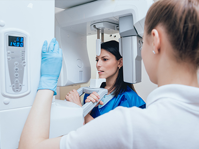
During a digital radiography appointment, a clinician or assistant will position a compact sensor inside the mouth for intraoral images or use an exterior device for panoramic views, depending on the exam. Exposures are brief and the sensor captures the image electronically, which appears on a monitor within seconds for immediate review. Most patients report minimal discomfort; the primary sensation is sensor placement rather than the exposure itself.
After the image is captured, the clinician will review it with the patient, using zoom and contrast tools to point out findings and explain recommendations. This visual review helps patients understand conditions and the rationale for treatment steps. If needed, additional views can be taken immediately to ensure complete diagnostic information.
Yes, digital radiographs are designed for secure electronic sharing, which facilitates referrals and collaborative care with specialists, laboratories and other providers. Files can be transferred using standard clinical formats and secure methods that preserve image quality and protect patient privacy. Quick sharing accelerates coordinated treatment planning and helps maintain continuity of care between multiple clinicians.
When transferring images, practices use secure channels and obtain any necessary patient consent consistent with privacy regulations. The ability to send full-resolution images reduces the need for repeat imaging when a patient seeks care elsewhere. Fast, reliable sharing supports timely consultations and efficient interdisciplinary collaboration.
Digital images are easy to archive, index and retrieve within the electronic dental record, which creates a continuous, accessible clinical history for each patient. This consistent record-keeping makes it simple to compare past and present images side by side to monitor cavities, bone levels, restorations and other changes over time. Reliable digital archives reduce the risk of lost films and help clinicians spot gradual trends that might otherwise be missed.
For patients with chronic conditions or complex restorative work, regular digital comparisons support timely interventions and more predictable outcomes. Digital storage also simplifies legal and clinical documentation by preserving high-quality images tied to the chart. At All About Smiles, digital records help maintain a cohesive diagnostic history that supports long-term oral health.
Yes, common digital radiograph types include bitewing, periapical and panoramic images, each serving distinct diagnostic roles. Bitewing images focus on the crowns of posterior teeth to detect interproximal decay and monitor bone levels between teeth, while periapical images capture the entire tooth and surrounding bone to evaluate roots and apical structures. Panoramic images provide a broad overview of the jaws, sinuses and temporomandibular joints for screening and planning purposes.
Advanced three-dimensional imaging, such as cone-beam computed tomography (CBCT), is available in some settings for detailed assessment of implant sites, impacted teeth and complex anatomy. Clinicians select the type of image based on diagnostic needs, balancing information gained against the appropriate level of exposure. Each radiograph plays a specific role in comprehensive evaluation and targeted treatment planning.
Clinicians minimize exposure by using high-sensitivity sensors, precise collimation, appropriate exposure settings and modern equipment that reduces scatter and unnecessary dose. They apply the principle of ALARA (as low as reasonably achievable) to ensure each image is justified and that the lowest practical exposure is used to obtain diagnostic quality. Proper patient positioning and technique reduce the likelihood of retakes, which helps limit overall radiation.
Selection of image type and frequency is tailored to each patient’s needs, with clinicians relying on professional guidelines and individual risk factors to determine appropriate intervals. When indicated, shielding and protective measures are used to protect sensitive areas. These combined practices ensure prudent radiation stewardship while maintaining diagnostic effectiveness.
Digital radiographs provide clear visuals that clinicians can display during consultations to explain findings and proposed treatments, making complex conditions easier to understand. Seeing an image on-screen helps patients visualize the issue and participate in informed decision-making, which improves clarity and alignment on next steps. The ability to annotate and enhance images supports focused discussions about specific concerns.
Clinicians often combine radiographs with intraoral photographs and digital treatment plans to illustrate options and expected outcomes. These visual tools create a collaborative environment where patients can ask questions and feel more confident about recommended care. At All About Smiles in Stockton, sharing digital images during visits is part of our approach to transparent, patient-centered communication.

Before or after office hours appointments available upon request.